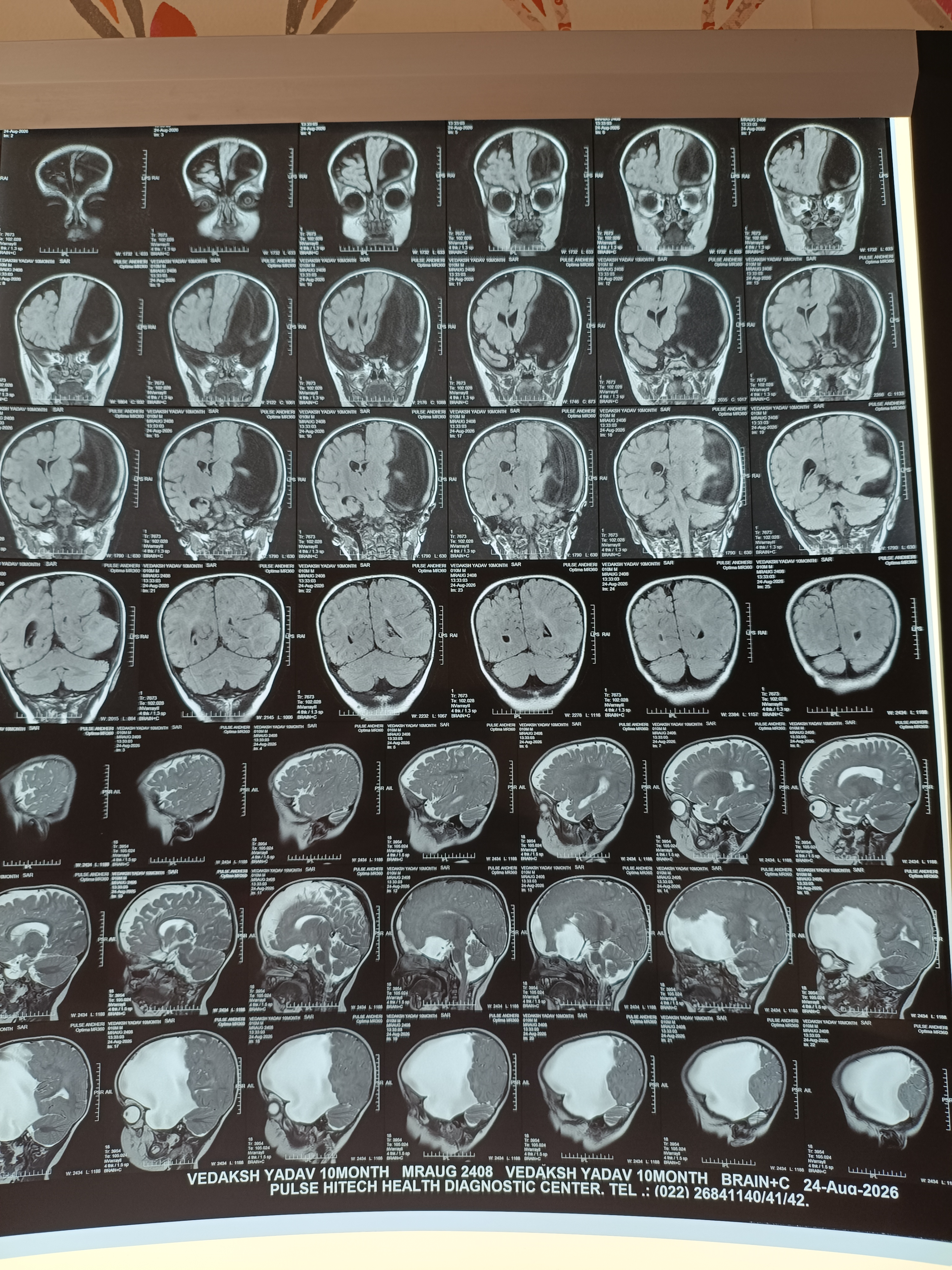
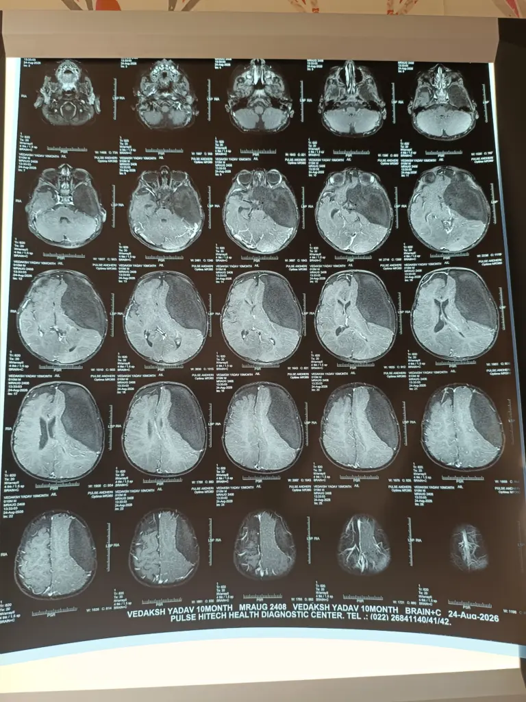
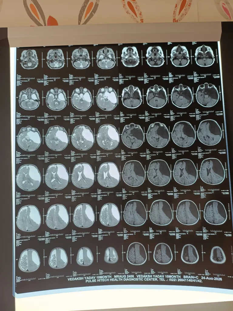
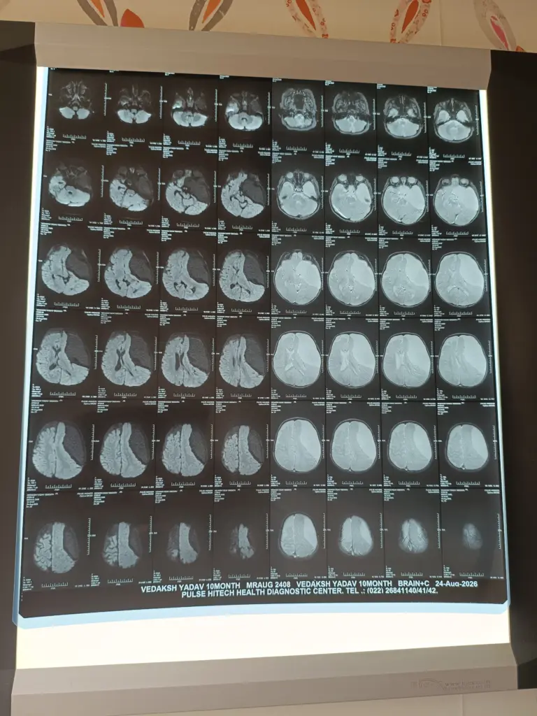
Evaluating an intracranial CSF-density cystic lesion in a young child requires precise differentiation, careful symptom correlation, and detailed neuroimaging analysis. Because these lesions behave fundamentally differently with respect to brain architecture and developmental potential, a structured approach is essential to avoid both unnecessary operations and delayed decompression.
Differential Diagnosis: Distinguishing Arachnoid Cysts, Porencephaly, and Schizencephaly
On routine or initial neuroimaging, these three entities can appear superficially similar because all contain clear, CSF-like fluid. However, their pathogenesis, relationship to brain parenchyma, and prognosis are completely distinct:
| Diagnostic Feature | Arachnoid Cyst | Porencephalic Cyst | Schizencephaly |
|---|---|---|---|
| Pathology / Origin | Congenital duplication/splitting of the arachnoid membrane with intra-cystic CSF accumulation. | Destructive, encephaloclastic lesion secondary to prior vascular insult, hemorrhage, or intrauterine infection. | Neuronal migration disorder resulting in a full-thickness cleft from the pial surface to the lateral ventricle. |
| Surrounding Parenchyma | Normal brain parenchyma, compressed or displaced by mass effect without intrinsic dysplasia. | Gliotic white matter lining the cyst wall (loss of preexisting brain tissue; ex vacuo expansion). | Cleft is characteristically lined by dysplastic, polymicrogyric gray matter. |
| Ventricular Communication | Non-communicating or valve-like ball-valve mechanism with basal cisterns/subarachnoid space; no direct ventricular breach. | Usually communicates directly with the adjacent ventricular system (ex vacuo porencephaly). | Directly extends from the ventricle to the cortical subarachnoid space (closed-lip or open-lip). |
| Mass Effect | Positive mass effect: Often expansile, scalloping adjacent bone, flattening gyri, shifting midline. | Negative / Neutral mass effect: Brain parenchyma is lost rather than compressed. | Structural gap: No mass effect unless an open-lip cleft leads to secondary CSF trapping/hydrocephalus. |
Clinical Presentation and Ongoing Symptomatology
Careful clinical correlation is paramount. Many arachnoid cysts are incidental discoveries on imaging obtained for unrelated minor head trauma or non-specific symptoms.
Key clinical features to track include:
- Signs of Raised Intracranial Pressure (ICP): Rapidly accelerating head circumference crossing percentiles, bulging or tense anterior fontanelle, persistent vomiting, sunsetting sign, or unexplained morning irritability and lethargy.
- Focal Neurological Deficits: Cranial nerve palsies (e.g., abducens nerve paresis), hemiparesis, or developmental regression/plateauing.
- Seizures: Unprovoked seizures or focal epilepsy, warranting correlation with EEG to verify if the epileptogenic focus directly matches the cyst margins.
- Endocrine / Visual Disturbance: Especially with suprasellar or middle fossa cysts causing compression of the optic apparatus or hypothalamo-pituitary axis (e.g., precocious puberty, developmental growth arrest).
High-Resolution Neuroimaging Evaluation
Static CT or standard MRI alone is often insufficient. High-resolution MRI (including thin-slice T2 sequences like CISS, FIESTA, or 3D-SPACE and phase-contrast CSF flow studies) is critical to answer three questions:
Internal Architecture & Communication
- Is the cyst completely isolated?
- Does it communicate with the native subarachnoid cisterns or deeper ventricular pathways?
- Is there a one-way (ball-valve) hydrodynamic trapping mechanism causing progressive fluid accumulation?
Dynamic Volumetric Change
- Is the cyst stable, or does sequential interval imaging demonstrate progressive enlargement over time?
Mass Effect on Critical Structures
- Degree of local parenchymal compression.
- Scalloping or remodeling of the overlying calvarium or skull base.
- Subcortical white matter effacement or distortion/obstruction of the ventricular outflow (resulting in secondary obstructive hydrocephalus).
Management Protocol: Conservative Surveillance vs. Surgical Intervention
Absolute Rule on Medical Management
- There is no medical therapy (pharmacotherapy, steroids, or diuresis) capable of resolving or shrinking an arachnoid cyst.
- The clinical decision is strictly binary: conservative serial observation versus surgical fenestration/decompression.
Indications for Intervention
Surgical intervention is indicated when there is:
- Progressive volumetric enlargement on sequential imaging.
- Significant, symptomatic mass effect with ventricular outflow distortion or midline shift.
- Intractable seizures refractory to medical therapy with concordant focal EEG findings.
- Neurological deficits, visual failure, or symptoms of raised ICP directly attributable to the lesion.
Asymptomatic, stable cysts without significant mass effect or hydrocephalus are safely managed with close clinical surveillance and spaced interval neuroimaging.
Surgical Approach: Endoscopic Marsupialization / Fenestration
When surgery is warranted, the treatment of choice is minimally invasive neuro-endoscopic marsupialization:
Operative Goal
- Transform an isolated, closed, pressurized fluid cavity into a freely communicating, unpressurized extension of the normal intracranial fluid pathways.
Technique
- Through a small burr hole or mini-craniotomy, a rigid neuro-endoscope is introduced into the cyst cavity.
- Rigid inspection of the cyst wall, vascular structures, and relation to adjacent cisterns/ventricles is performed.
- Wide, multiple fenestrations (marsupialization) are created between the cyst and the deeper basal cisterns (e.g., prepontine, interpeduncular, or chiasmatic cisterns) or into the ventricular system (ventriculocystostomy).
Advantages over Shunting
- Avoids the lifelong hardware dependence, occlusion risks, mechanical failures, and infection rates associated with cystoperitoneal (CP) shunts.
- Restores physiological, internal CSF flow dynamics within native intracranial compartments.
This page is for general education only and does not replace an in-person consultation. If your child shows sudden changes — seizures, persistent vomiting, drowsiness or wound leakage — go to the nearest emergency department immediately.